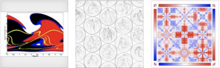
Images depicting star collisions, atom movement in yeast ribosomes, and herbaceous plant root scans were the winning entries of the 2020 Wesleyan Scientific Imaging Contest.
The Wesleyan Scientific Imaging Contest, held annually in August, recognizes student-submitted images—from experiments or simulations done with a Wesleyan faculty member—that are scientifically intriguing, as well as aesthetically pleasing. The contest is organized by the College of Integrative Sciences as part of the summer research program.
The winners included Osama Elgabori ’22, Carol Dalgarno ’21, and Jolie Villegas ’21. Elgabori’s advisor is Brian Stewart, professor of physics; Dalgarno’s advisor is Michael Weir, professor of biology; and Villegas’ advisor is Sonia Sultan, professor of biology.
Three faculty members served as judges: Brian Northrop, associate professor of chemistry; Amy MacQueen, associate professor of molecular biology and biochemistry; and Meng-ju (Renee) Sher, assistant professor of physics.
“What we look for is both the art and the science,” Sher said. “Most of the top candidates have works that intrigued us and drew us in to find out more. Once our curiosity is up, we learn the science behind this image from the descriptions students submit. For me, learning science from the art piece is what led me to identify these three winners.”
This year, 12 students submitted contest entries, which due to the COVID-19 pandemic, was more than the judges had expected.
“Typically we receive many microscopic images or photographs students took in the lab. Without a physical research lab for most of our undergraduate students, we were very impressed with the type of images they put together,” Sher said.
The first-place winner (Elgabori) received a $150 prize, the second-place winner (Dalgarno) received $100, and the third-place winner (Villegas) received $50. Prizes were funded by the Office of Academic Affairs.
The winning images are shown below, along with scientific descriptions written by the students:
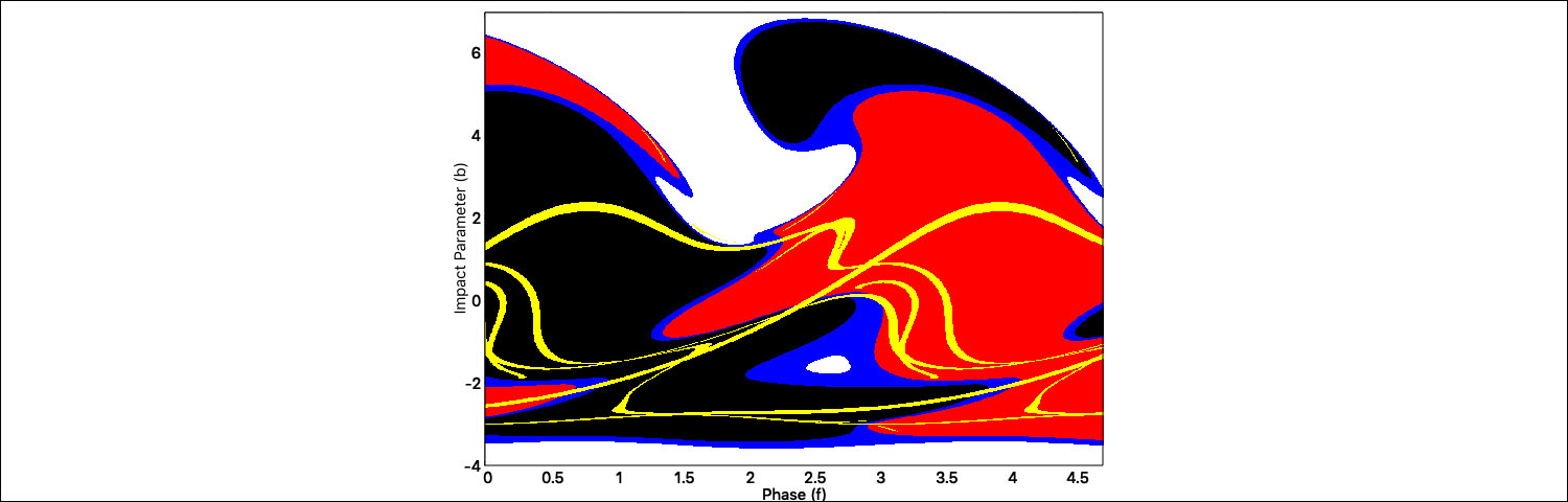
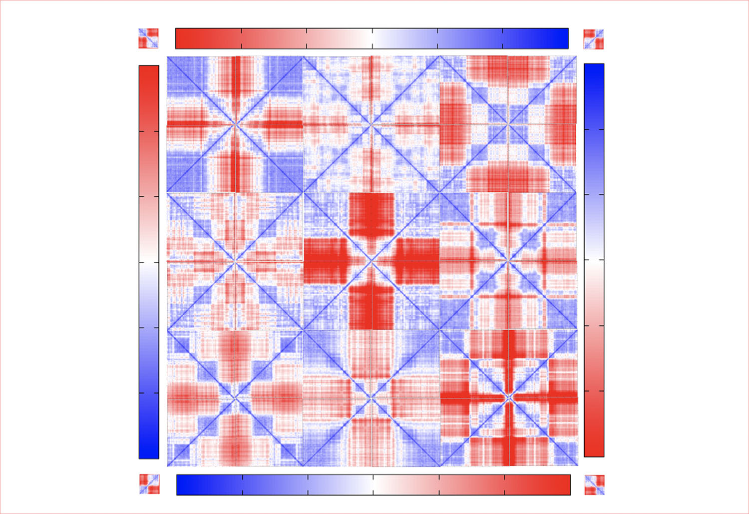
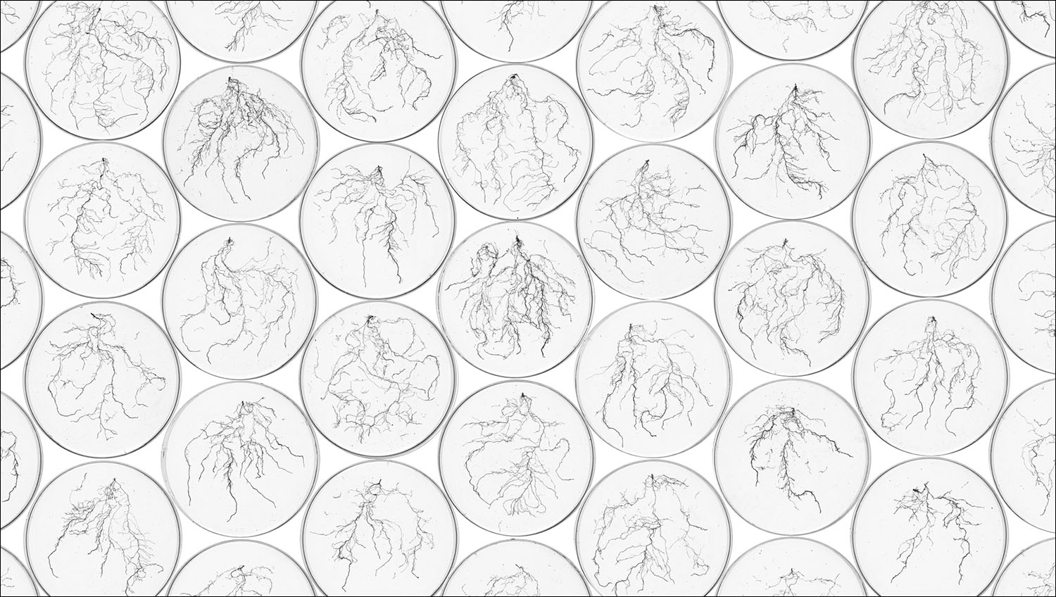
Osama Elgabori ’22 took first place with his image of a color grid for a million orbit numerical scattering experiment of three-body collisions between a binary star and field star. “The white space indicates fly-by events in which the field star misses the binary star, leaving the binary intact with a new orbit. The black and red points represent exchanges with mass 1 or mass 2 respectively, in which the field star takes the place of one the members in the original binary star and forms a new binary. Blue points represent dissociation events in which the binary dissolves and all three stars leave in different directions. Yellow points are trajectories that failed to conserve total energy to within 1% of the initial set up of the system.”Carol Dalgarno ’21 took second place with her image titled “Quilting Conformational Changes.” “Molecular dynamics (MD) computer simulations allow us to study conformational changes in biological systems, producing a trajectory of snapshot frames that capture the movement of atoms over time. Using RMS2D calculations we can calculate the root mean square deviation (RMSD) between the atom positions of two frames in a trajectory. The output of the RMS2D function gives a matrix of all possible frame-to-frame RMSD calculations for a single MD trajectory. These matrices are plotted as heat maps where red indicates less similar structures (higher RMSD) and blue indicates more similar structures (lower RMSD). This image shows RMS2D heatmaps of ten 50 nanosecond MD simulations of the decoding center of the yeast ribosome with each heat map copied and rotated three times. Each simulation has its own distinct pattern of red and blue blocks, suggesting that the system samples many different transiently stable conformations over the course of the trajectory.”Jolie Villegas ’21 took third place with her image of Polygonum cespitosum root scans. (The herbaceous plant also is known as tufted knotweed or oriental lady’s thumb.) “Rather than submit a digital image of a single root, I created a collage to show the beauty in diversity that exists within populations and life in general. While these plants appear so simple and similar above ground, their complex root systems reveal that there is so much more to life beneath the surface.”