Wesleyan Students Recognized for Scientific Images

Lauren RubensteinAugust 8, 20167min
carter

This summer, Stephen Devoto, professor of biology, professor of neuroscience and behavior, launched the inaugural Wesleyan Scientific Imaging Contest. The contest, which recognizes student-submitted images from experiments or simulations done with a Wesleyan faculty member that are scientifically intriguing as well as aesthetically pleasing, drew 35 submissions from the fields of physics, biology, molecular biology and biochemistry, psychology, earth and environmental science, chemistry and astronomy.

Participants submitted an image along with a brief description written for a broad, scientifically literate audience. The entries were judged based on the quality of the image and the explanation of the underlying science. The first-place prize went to Eliza Carter ’18 from the Earth and Environmental Science Department. Aidan Stone ’17 and Jeremy Auerbach ’17 tied for second places, while Riordan Abrams ’17 won third place. The images were judged by a panel of four faculty members: Devoto; Ruth Johnson, assistant professor of biology, assistant professor of integrative sciences; Brian Northrop, assistant professor of chemistry, assistant professor of integrative sciences; and Candice Etson, assistant professor of physics.

The first-place winner receives a $200 prize; the second-place winner receives $100; and the third-place winner receives $50. Prizes were funded by the Office of Academic Affairs.

Devoto was inspired by a similar contest that his daughter won at Haverford College.

“Students at Wesleyan produce extraordinary scientific images, ranging from graphs and computer simulations to microscope and telescope images,” he said. “I wanted students to have fun, to think of their scientific images in an artistic sense. And I thought that the artistic presentations of student scientific images would be a striking testament to the quality and fun of student research here. I hope these will be displayed on campus to highlight the science and the creativity, which thrive at Wesleyan.”

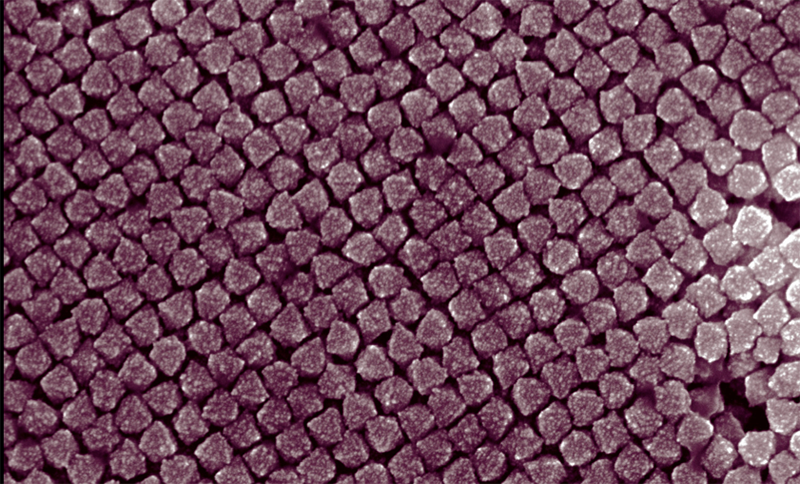
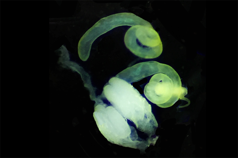
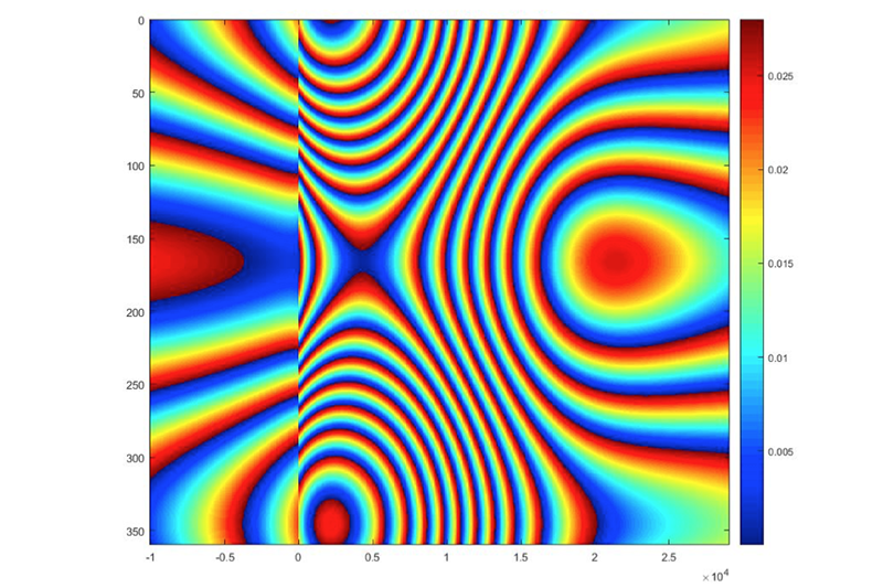
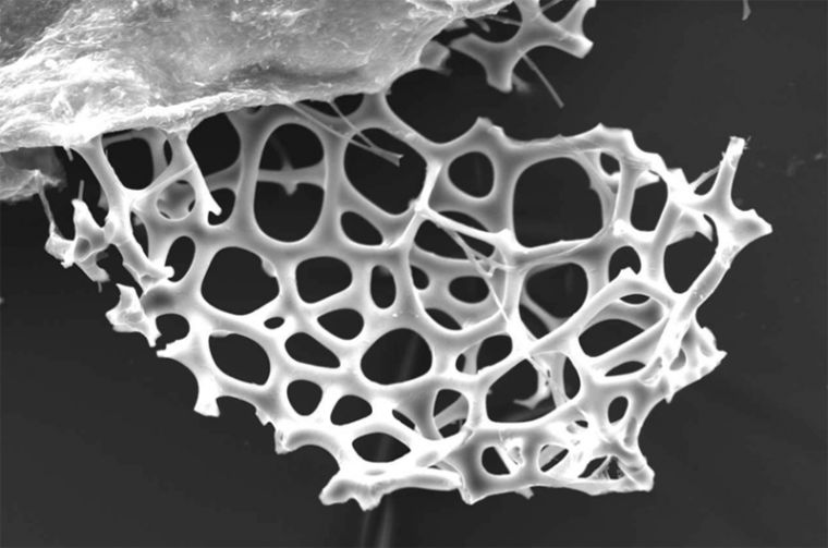
The four winning images are shown below, along with scientific descriptions:

Eliza Carter '18 submitted a scanning electron microscope image of the shell of a radiolarian (a protozoa) found near the top of an Antarctic sediment core from ODP site 697. The radiolarian shell is around 2.7 million years old and is made from silica that was produced by the radiolarian. Studying the percent biogenic silica in a sediment sample is a proxy for primary productivity: the more biosilica you have, the more productive it was.
Eliza Carter ’18 submitted a scanning electron microscope image of the shell of a radiolarian (a protozoa) found near the top of an Antarctic sediment core from ODP site 697. The radiolarian shell is around 2.7 million years old and is made from silica that was produced by the radiolarian. Studying the percent biogenic silica in a sediment sample is a proxy for primary productivity: the more biosilica you have, the more productive it was.

Aidan Stone '17 submitted a scanning electron microscope photograph of the fuzzy surface structure of platinum nanoparticles. The nanoparticles, which are about 70 nanometers in diameter, were synthesized with silver additive, which promotes the formation of platinum-silver alloys. Certain highly faceted structures make platinum nanoparticles useful as catalysts in industrial chemical reactions.
Aidan Stone ’17 submitted a scanning electron microscope photograph of the fuzzy surface structure of platinum nanoparticles. The nanoparticles, which are about 70 nanometers in diameter, were synthesized with silver additive, which promotes the formation of platinum-silver alloys. Certain highly faceted structures make platinum nanoparticles useful as catalysts in industrial chemical reactions.
Jeremy Auerbach '17 submitted an image depicting a pair of spiral-shaped testes isolated from a single adult Drosophila melanogaster fly, as well as a pair of ovaries isolated from a single adult female of the same species. Drosophila melanogaster testes are unique in their radiant yellow color. Each ovary consists of several tubular egg-producing ovarioles, which are visible in the image, and a transparent oviduct connects the two ovaries. The striking glow of these reproductive tissues is entirely natural.
Jeremy Auerbach ’17 submitted an image depicting a pair of spiral-shaped testes isolated from a single adult Drosophila melanogaster (common fruit fly), as well as a pair of ovaries isolated from a single adult female of the same species. Drosophila melanogaster testes are unique in their radiant yellow color. Each ovary consists of several tubular egg-producing ovarioles, which are visible in the image, and a transparent oviduct connects the two ovaries. The striking glow of these reproductive tissues is entirely natural.
Riordan Abrams '17 submitted an image that shows deformities in the ground caused by an earthquake. This 2D computer model of Synthetic Aperture Radar data demonstrates how the north-south orientation of the fault affects the deformation pattern observed in an interferogram. This occurs because the angle at which the satellite is viewing the deformation field impacts what it sees.
Riordan Abrams ’17 submitted an image that shows deformities in the ground caused by an earthquake. This 2D computer model of Synthetic Aperture Radar data demonstrates how the north-south orientation of the fault affects the deformation pattern observed in an interferogram. This occurs because the angle at which the satellite is viewing the deformation field impacts what it sees.