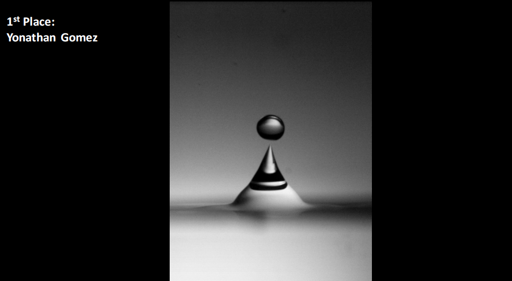
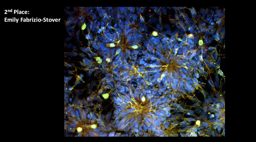
Plasma Bubble, Stem Cell Images Win Scientific Imaging Contest

Olivia DrakeAugust 11, 20175min
graph

This summer, Wesleyan hosted the second annual Wesleyan Scientific Imaging Contest, which recognizes student-submitted images from experiments or simulations done with a Wesleyan faculty member that are scientifically intriguing as well as aesthetically pleasing. This year, 33 images were submitted from six departments.

The entries were judged based on the quality of the image and the explanation of the underlying science.  The images were judged by a panel of four faculty members: Steven Devoto, professor of biology, professor of neuroscience and behavior; Ruth Johnson, assistant professor of biology, assistant professor of integrative sciences; Brian Northrop, assistant professor of chemistry, assistant professor of integrative sciences; and Candice Etson, assistant professor of physics.

The first-place winner received a $200 prize; the second-place winner received $100; and the third-place winner received $50. Prizes were funded by the Office of Academic Affairs.

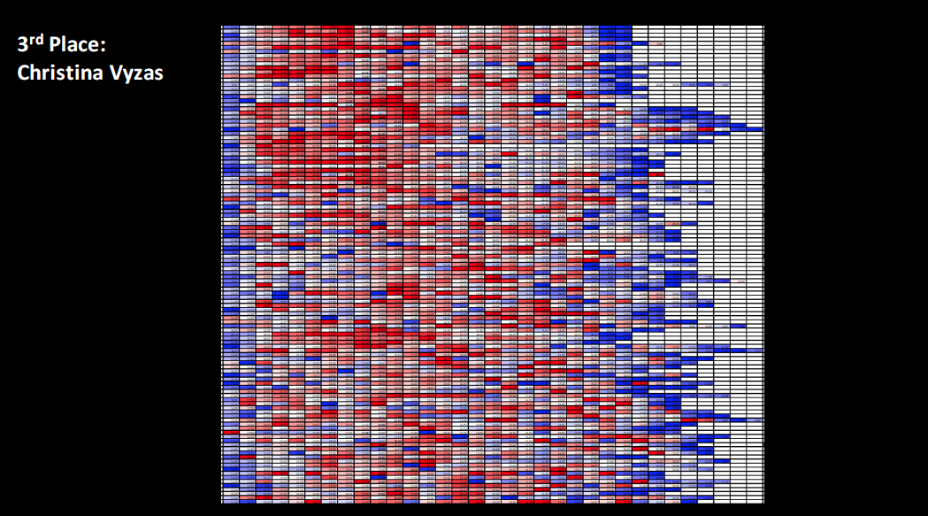
The three winning images are shown below, along with scientific descriptions, written by the students.

Yonathan Gomez '18 won first place with his image, "Jumping" Drop. The drop is an expanding partially-ionized plasma created underwater by a pulsed Nd:YAG laser, which pushes upwards on the surface of the water. As the plasma bubble expands, it disrupts the surface from below, which launches a water drop upward. The water drop shown has a diameter of approximately 2mm. The image was taken at 1/2,000 frames per second.
Yonathan Gomez ’18 won first place with his image, “Jumping” Drop. The drop is an expanding partially-ionized plasma created underwater by a pulsed Nd:YAG laser, which pushes upwards on the surface of the water. As the plasma bubble expands, it disrupts the surface from below, which launches a water drop upward. The water drop shown has a diameter of approximately 2mm. The image was taken at 1/2,000 of a second.

Emily Fabrizio-Stover Embryonic stem cells differentiating into neurons in vitro from neural rosettes, a structure of radially arranged neural stem cells. Because these structures share properties with the neural tube, they can be used to model neurogenesis. Immunocytochemistry highlights the nuclei in blue (Hoechst) and cells in mitosis in yellow (Phospho-histone H3 in green and Numb in red).
Emily Fabrizio-Stover ’18 won second place with her image of embryonic stem cells differentiating into neurons in vitro from neural rosettes, a structure of radially arranged neural stem cells. Because these structures share properties with the neural tube, they can be used to model neurogenesis. Immunocytochemistry highlights the nuclei in blue (Hoechst) and cells in mitosis in yellow (Phospho-histone H3 in green and Numb in red).
Christina Vyzas '18 took third place with this image of a portion of a chart conditionally formatted for zebrafish centra size range. The scale from blue to red indicates the variation from smallest to largest centra down the length of the vertebral column in the posterior direction.
Christina Vyzas ’18 took third place with this image of a portion of a chart conditionally formatted for zebrafish centra size range. The scale from blue to red indicates the variation from smallest to largest centra down the length of the vertebral column in the posterior direction.